Amniotic Fluid Stem Cells: New Hopes for Organoid Research
How Italian-led scientific research is turning cells collected from amniotic fluid into laboratory-grown organoids, opening new ways to investigate fetal development and human tissue biology.
Stem cell research continues to expand the ways scientists study the development and organization of human tissues. In an important research advance involving an Italian-led international collaboration, cells recovered from amniotic fluid were used to generate three-dimensional organoids resembling developing fetal tissues. The work highlights the scientific value of amniotic fluid as a source of cells for investigating organ development and building more representative laboratory models.
The study, published in Nature Medicine in 2024 and reported by Nature Italy, investigated epithelial cells present in amniotic fluid and their ability to form organoids with characteristics associated with the lungs, intestines and kidneys. This approach could support research into how fetal organs develop and how selected biological processes can be studied outside the body.
1. Understanding the Italian Stem Cell Research Advance
Traditional prenatal investigations often rely on imaging techniques and genetic analyses. These methods provide important information, but they do not always reveal how a developing organ functions at a cellular or tissue level. Researchers are therefore investigating complementary laboratory models that can help explain the biology behind observations made during pregnancy.
The Italian-led study explored whether epithelial cells present in amniotic fluid could be identified, characterized and cultivated to produce organoids. Rather than relying exclusively on tissue samples from an already developed organ, the research used cells associated with fetal development that were present in fluid collected during prenatal procedures.
The researchers characterized cellular populations using single-cell analysis and investigated whether selected cells could grow into organized three-dimensional structures. The resulting organoids displayed features associated with their tissue of origin, creating opportunities for further developmental biology research.
Why this research matters
Amniotic fluid may contain cells originating from different developing fetal tissues. Identifying these populations and growing suitable cells as organoids could give researchers additional models for studying organ development, tissue organization and selected biological processes.
2. What Is Amniotic Fluid?

Amniotic fluid surrounds the developing fetus during pregnancy. It supports fetal development and contains a mixture of biological materials, including cells and cellular products originating from different fetal tissues. Its cellular composition can provide valuable information for developmental biology research.
During certain prenatal procedures, samples of amniotic fluid are collected for established clinical purposes. Depending on the sample and applicable ethical and regulatory requirements, researchers may investigate cells in these samples to understand their identity, properties and potential uses in laboratory models.
Importantly, amniotic fluid is not a uniform population of stem cells. It contains different cell types, and each population must be carefully characterized. Scientists need to determine which cells are present, whether they are viable and what biological characteristics they retain under laboratory culture conditions.
3. What Are Amniotic Fluid Stem Cells?
Amniotic fluid stem cells are a broad area of stem cell biology research. The term is commonly used when discussing stem or progenitor cell populations isolated from amniotic fluid. These populations are biologically diverse, and their properties depend on the specific cells, isolation method and experimental conditions being studied.
Stem cells are studied for their ability to self-renew and, depending on the cell population, differentiate into particular specialized cell types. Progenitor cells can also generate selected cell lineages, but generally have a more restricted developmental potential than broadly pluripotent stem cells.
Cellular diversity
Amniotic fluid contains cells with different origins and biological characteristics, making cellular identification an important first step in research.
Cell characterization
Researchers use molecular markers, microscopy and gene-expression analysis to identify cell populations and investigate their properties.
Three-dimensional culture
Selected cells can be investigated in culture systems that support the formation of organized structures resembling aspects of tissues.
A key distinction is that the cells used to generate organoids in the 2024 study included fetal epithelial stem or progenitor populations. This is not the same as claiming that every cell in amniotic fluid is a stem cell or that all amniotic fluid-derived cells can form every type of organoid.
4. How Are Organoids Developed From Amniotic Fluid Cells?
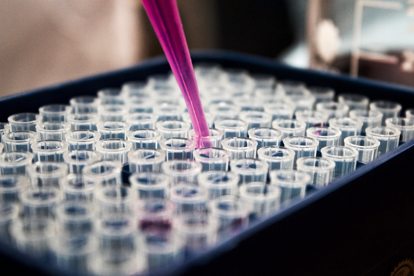
Organoids are three-dimensional, laboratory-grown cellular structures that reproduce selected structural or functional characteristics of organs. They are not complete miniature organs. Instead, they model particular aspects of tissue organization and biology that researchers can examine under controlled conditions.
In amniotic fluid organoid research, the general workflow involves identifying relevant cells, isolating suitable populations, expanding them under appropriate culture conditions and assessing the structures that develop. The specific procedures depend on the cell type and intended research question.
Cell identification
Single-cell analysis and other characterization methods help researchers identify cellular populations and investigate their potential tissue origins.
Cell isolation and culture
Suitable cells are selected and cultured under conditions designed to support their survival, expansion and organization.
Organoid characterization
Microscopy, gene-expression profiling and protein analysis can establish whether the resulting structures exhibit characteristics associated with particular tissues.
Researchers can then compare organoid structure, cellular composition and molecular features with relevant reference tissues or other experimental models. Such comparisons help establish what an organoid can reliably represent and where its limitations remain.
5. Lung, Intestinal and Kidney Organoids: Three Research Models
The 2024 study reported organoids with characteristics associated with multiple developing fetal tissues. These models illustrate how cells obtained from a single type of biological sample may provide access to different research systems.
| Organoid model | Research focus | Potential scientific value |
|---|---|---|
| Lung organoids | Developing pulmonary epithelium and tissue organization | Investigating selected aspects of lung development and fetal tissue biology. |
| Intestinal organoids | Developing gastrointestinal epithelial structures | Studying intestinal cell organization, differentiation and molecular characteristics. |
| Kidney organoids | Cellular characteristics associated with developing renal tissue | Exploring aspects of kidney development and tissue-specific biology. |
Lung organoids and fetal development
Lung organoids can provide a controlled environment for studying selected characteristics of developing pulmonary tissue. In the Italian-led research, the team also generated lung organoids from samples associated with a fetus with congenital diaphragmatic hernia. This demonstrated the potential to investigate selected features of a specific developmental condition in a laboratory model.
Intestinal organoids and epithelial biology
Intestinal organoid models allow researchers to investigate epithelial organization and cellular differentiation. Their value depends on which intestinal features are reproduced and how closely the model reflects the biological process being studied.
Kidney organoids and renal tissue research
Kidney-associated organoids offer another way to explore the organization and molecular identity of developing renal cells. Additional validation is necessary before conclusions from an organoid can be generalized to a complete kidney or its full physiological functions.
6. Potential Applications of Amniotic Fluid Organoid Research
The significance of this research extends beyond the creation of three-dimensional cell cultures. Carefully characterized organoids could provide experimental systems for studying biological processes that are difficult to observe directly during human fetal development.
Developmental biology research
Organoids may help researchers examine how selected fetal epithelial cells organize, differentiate and develop tissue-specific features. This can complement genetic analysis, imaging and other laboratory techniques used to investigate early human development.
Cellular and molecular analysis
Researchers can study gene expression, protein localization and cellular morphology within organoids. Combining microscopy with molecular biology methods may reveal differences between cell populations, developmental stages and experimental conditions.
Laboratory models for specific biological conditions
Organoids derived from selected samples may help investigate how particular developmental abnormalities affect cellular characteristics. However, an organoid represents only selected aspects of the original tissue and does not reproduce every biological feature of a fetus.
Experimental testing and model evaluation
In some research settings, organoids can be used to investigate cellular responses to experimental compounds or changes in culture conditions. Such findings are preliminary and model-dependent. Results from an organoid must not automatically be interpreted as evidence of safety, effectiveness or outcomes in humans.
Personalized research models
Where suitable samples and validated protocols are available, sample-specific cellular models may help scientists explore biological variation between individuals. Further work is needed to determine when these approaches provide reproducible and clinically meaningful information.
7. Why Are Researchers Interested in Amniotic Fluid as a Cell Source?
Amniotic fluid is of interest because it can contain cells associated with multiple developing tissues. In appropriate research settings, samples collected during clinically indicated prenatal procedures may provide an opportunity to investigate these cells without obtaining a separate tissue biopsy from the developing organ.
Access to developing cells
Samples can contain cellular populations that provide insights into fetal tissue development.
Multiple tissue models
Different identified cell populations may support the development of organoids representing different tissue types.
Complementary methods
Organoids can be investigated alongside sequencing, microscopy, proteomics and other molecular research techniques.
These opportunities do not mean that amniotic fluid sampling is risk-free, that samples are always suitable for organoid culture or that an experimental model is ready for clinical use. The original reason for sample collection, informed consent, sample handling and research oversight remain essential considerations.
8. Scientific Challenges and Limitations
Although the findings are promising, amniotic fluid-derived organoid research remains an evolving field. Several questions must be addressed before these models can be used reliably across broader research settings.
Cellular variability
The number and types of cells in amniotic fluid can vary between samples. Differences in gestational stage, sample collection and processing may affect the cell populations available for culture.
Reproducibility of organoid culture
Researchers need robust protocols that produce consistent organoids across experiments. Culture conditions, cell selection and characterization criteria can all influence the results.
Model fidelity
An organoid reproduces selected characteristics of a tissue rather than the entire structure and function of a complete organ. Scientists must establish which biological features are represented and which are absent.
Ethical oversight and sample consent
Research involving human prenatal samples requires appropriate informed consent, ethical review, privacy safeguards and compliance with applicable laws and institutional policies. Sample availability does not by itself establish permission for every possible research use.
Translation beyond laboratory research
Additional validation is needed before these models can support clinical decisions or be used to predict outcomes for individual pregnancies. Laboratory findings should be interpreted within the limits of the study design and evidence available.
9. The Future of Stem Cell and Organoid Research

Future research may refine the methods used to identify fetal epithelial cell populations, expand selected cells and establish reproducible organoid cultures. Improved molecular characterization could also help scientists compare organoids with reference tissues and understand how accurately each model represents its tissue of origin.
Combining single-cell sequencing with organoids
Single-cell RNA sequencing can help characterize gene-expression patterns across individual cells. When combined with organoid culture, it may enable researchers to investigate cellular diversity, identify distinct populations and examine how gene-expression profiles change during experimental development.
Integrating imaging and molecular biology
Microscopy, immunostaining, transcriptomic analysis and protein characterization can provide complementary information about organoid structure and identity. Integrating these methods may improve the evaluation of tissue-specific features and experimental reproducibility.
Building more representative human tissue models
One longer-term objective in organoid research is to create models that better reproduce selected aspects of human biology. Achieving this requires careful validation, transparent reporting and a clear understanding of each model's limitations.
The Italian-led amniotic fluid study adds to this growing area of research by demonstrating that cells associated with developing fetal tissues can be used to generate several types of organoids. Further studies will determine how broadly these models can be applied and what information they can reliably provide.
10. Frequently Asked Questions About Amniotic Fluid Stem Cells
What are amniotic fluid stem cells?
The term describes stem or progenitor cell populations studied from amniotic fluid. The fluid contains different cell types, so individual populations must be identified and characterized rather than treating every cell as a stem cell.
What did the Italian-led study discover?
Published in 2024, the research demonstrated that selected epithelial cells from amniotic fluid could be cultivated into organoids with characteristics associated with developing lungs, intestines and kidneys.
What are amniotic fluid-derived organoids?
They are three-dimensional laboratory-grown cellular structures generated from selected cells in amniotic fluid. They reproduce certain features of developing tissues but are not complete organs.
Which organs were represented in the research?
The study reported organoids associated with lung, intestinal and kidney tissues. Each model represents selected biological features and requires appropriate validation.
Can organoids help scientists study fetal development?
They may complement other research methods by allowing selected cellular and molecular processes to be investigated in controlled laboratory conditions. Their usefulness depends on the model and the question being studied.
Are amniotic fluid stem cell organoids already routine clinical tools?
No. The research approach is promising, but further validation is needed before such models can be considered established clinical tools for individual prenatal assessments.
How does single-cell RNA sequencing contribute to organoid research?
Single-cell RNA sequencing measures gene-expression patterns in individual cells. It can help identify cellular populations and compare their molecular profiles with those of cultured organoids or reference tissues.
What are the main limitations of organoid models?
Organoids do not reproduce every cell type, structural feature or physiological function of a complete organ. Variability between samples and culture protocols can also affect reproducibility.
Scientific References and Further Reading
The following scientific sources provide background on amniotic fluid cells, fetal organoids and stem cell research.
- Nature Italy — Growing organoids from amniotic fluid may help prenatal diagnosis (2024)
- ANSA — Miniature organs obtained from amniotic fluid stem cells
- PubMed — Characterization of fetal human amniotic fluid cells using single-cell analysis (2023)
- Biomedicines — Research on fetal human amniotic fluid cells